A pelvic ultrasound is a non-invasive diagnostic exam that allows quick visualisation of the organs and structures within the pelvis. It is usually used to assess a woman’s uterus, cervix, vagina, fallopian tubes, and ovaries.
A pelvic ultrasound may be used to diagnose abnormalities such as tumours or for evaluating fertility and monitoring foetal development.
Although ultrasound is a valuable diagnostic tool, it cannot give a definite diagnosis of cancer or specific disease.
Why Would You Need A Pelvic Ultrasound?
A pelvic ultrasound has a few different applications. It may be used for measuring or evaluating your pelvic organs or for diagnosing and treating gynaecological abnormalities. It is a valuable tool to assist with other procedures such as endometrial biopsy.
When an ultrasound is performed to measure and evaluate your pelvic organs, information such as the size, shape, thickness, and position of your organs is recorded. Blood flow through the pelvic organs can also be observed. Abnormalities, such as the presence of fluids or masses in the pelvic cavity can be observed.
Conditions That Can Be Diagnosed And Treated
Pelvic ultrasound may be used to diagnose and assist in the treatment of the following conditions:
- Abnormalities in the structure of the uterus, such as endometrial conditions
- Fibroids (benign growths), masses and cysts
- To locate the position of an intrauterine contraceptive device (IUD)
- Signs of inflammation or infection such as pelvic inflammatory disease (PID)
- Postmenopausal bleeding
- Monitoring of ovarian follicle size to evaluate infertility
- Aspiration of eggs from ovaries for in vitro fertilisation
- Ectopic pregnancy (pregnancy occurring outside of the uterus)
- Monitoring foetal development during pregnancy
- Assessing certain foetal conditions
Ultrasound During Pregnancy
You will normally have at least 2 ultrasound scans during your pregnancy - at 10 to 14 weeks and between 18 and 21 weeks. You may need to have more than 2 scans, depending on your health and the pregnancy.
The first ultrasound scan is also known as the dating scan. From this scan the estimated date of delivery (EDD) can be predicted based on the baby's measurements. This scan can include a nuchal translucency (NT) scan, which screens for Down's syndrome.
The second mid-pregnancy scan usually takes place between 18 and 21 weeks of pregnancy. This scan checks for 11 physical conditions in your baby. These include conditions such as open spina bifida, cleft palate and serious cardiac abnormalities.
An ultrasound will determine if you are having more than 1 baby and will show the position of your baby and the placenta. If the placenta is low down in late pregnancy, a caesarean section may be advised.
How Does Ultrasound Work?
Ultrasounds use a transducer that sends out ultrasound waves at a high frequency. When the ultrasounds transducer is placed on the abdomen or in the vagina, the ultrasound waves move through the pelvic organs and structures. The transducer processes sound waves which are converted into an image of the organs or tissues being examined. No radiation is used during the procedure.
Certain factors or conditions may interfere with the results of the test. These include:
- Severe obesity
- Barium within the intestines (from a recent barium procedure)
- Intestinal gas
- An empty bladder
How Is A Pelvic Ultrasound Performed?
It is best to have a full bladder when the scan is done. You should not empty your bladder until after the exam. Besides feeling some pressure from having a full bladder, you should not feel too much discomfort during the scan.
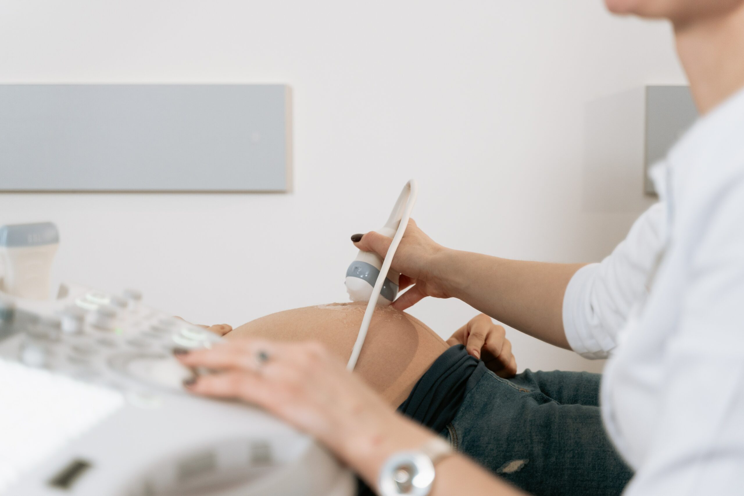
Pelvic ultrasound can be performed in one of two ways - either through your abdomen or through your vagina.
Transabdominal Scan
This scan is done through your abdomen. A transducer is placed on the abdomen using conductive gel and then gently moved over your abdomen.
Transvaginal Scan
This scan is done through your vagina. A long, thin lubricated transducer probe is inserted into your vagina. The probe is covered with a lubricated condom. The probe may be moved around to get a better view which may cause a small amount of discomfort but it should not be painful.
During the scan, you will lie down on the examination table on your back. For a transvaginal scan, you will need to keep your knees bent during the scan. For both types of scans, the images are shown on a video monitor.

 71–75 Shelton Street, Covent Garden, London, WC2H 9JQ
 +44 (0) 20 4577 1127

